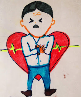
Approach to a case of chest pain
Approach to a case of chest pain
Initial diagnostic assessment and triage is made around three categories: -
a) Myocardial Infarction
b) Cardiopulmonary causes
1) Pericardial diseases
2)Aortic emergencies
3) Pulmonary conditions
c) Non cardiopulmonary causes
Most common cause- Gastrointestinal diseases
Causes of chest discomfort
- Gastrointestinal (oesophageal reflux, oesophageal spasm, peptic ulcer disease)
- Ischemic heart disease
- Chest wall syndrome
- Pericarditis
- Pleuritis
- Pulmonary embolism
- Lung cancer
- Aortic aneurysm
- Aortic stenosis
- Herpes zoster
- Costrochondritis
- Cervical rib
- Trauma
How to Approach ?
History
In case of non traumatic chest pain
Pain-Quality, Location and radiation, Pattern(onset and duration) , Provocative and alleviating factors ,associated symptoms.
Quality of pain
- Pressure or tightness-MI(some patients may deny any pain and some may have only dyspnoea or vague anxiety)
- Steady ,severe pressure or aching-Massive pulmonary embolism, Pericarditis
- Tearing or ripping -Acute aortic dissection
- Sharp stabbing-Pleuritic chest pain
- Burning type-Acid reflux , Peptic Ulcer disease, Esophageal pain with spasm
Location of pain
- MI-Substernal pain radiating to neck, jaw, shoulder, arms.
- Oesophageal pain-retrosternal
- Aortic dissection-Severe pain radiating to back between shoulder blades
- Pericardial pain radiates to trapezial ridge
Pattern of pain
- MI-builds in minutes, increased on activity and decreases on taking rest
- Aortic dissection, Pulmonary embolism, Spontaneous pneumothorax-Pain reaches peak intensity immediately
- Fleeting pain-for only few seconds
Provocative and Alleviating factors
- MI- decreases on rest ( except warm up angina)
- Musculoskeletal- Altered intensity with changes in position
- Pericarditis-worse in supine and sitting position but decreases on leaning forward and upright position
- GE reflux-Aggravated by alcohol, some food or reclined position
Pain increased on eating- Peptic ulcer disease, Acute pancreatitis and cholecystitis, Coronary artery disease (due to redistribution to splanchic vasculature)
Associated symptoms
- MI-Dyspnoea, diaphoresis, nausea ,fatigue, faintness, syncope, pre-syncope etc.
- Pulmonary embolism-Haemoptysis, syncope
- Severe heart failure-blood tinged frothy sputum
- Nausea and vomiting-MI(activation of vagal reflex)
Past Medical History
Physical Examination
General
Patient-Anxious, uncomfortable, pale, cyanotic, diaphoretic-MI, Acute cardiopulmonary disorder
Levine's Sign
Vitals
Tachycardia, hypotension-Acute MI, Cardiigenic Shock, Pericarditis with Tamponade, Massive pulmonary embolism, Tension pneumothorax
Severe Hypertension or hypotension-Aortic emergencies
Sinus Tachycardia-Submassive Pulmonary embolism
Tachypnoea and hypoxemia-Pulmonary cause
Systemic Examination
Pulmonary System-Findings can localise a primary pulmonary cause of chest discomfort like asthma, pneumothorax, pneumonia
Cardiac-S3 or S4 heared in myocardial dysfunction
Cardiac murmers and Pericardial rub
Abdominal - Localised tenderness
Right Ventricular Dysfunction-Hepatic congestion
Vascular
Pulse deficit-Chronic Atherosclerosis
Aortic dissection leads to acute limb ischemia with loss of pulse and pallor
Musculoskeletal System
-Pain arising from costrochondral and chondrosternal articulation is associated with localized swelling ,redness, tenderness.
Investigations
ECG
Chest X ray
Cardiac Biomarkers
CT angiography


Comments
Post a Comment